Lesson: MRI Safety Challenge
 (Lesson courtesy of TeachEngineering and VU Bioengineering RET Program, School of Engineering, Vanderbilt University.) Grade Level: 11-12. Time Required: 2-3 class periods.
MRI Safety Challenge
Summary
In this lesson, students in grades 11 and 12 think like a safety engineer to question the risks and potential dangers associated with magnetic resonance imaging, commonly used today to produce images of soft tissues and bones. Students write journal responses and brainstorm about the kind of information they need to answer the question of MRI safety. They share their ideas with the class, then watch a video interview with a researcher to gain a professional perspective on MRI safety and brainstorm any additional ideas. Drawing together what they have learned, they produce a pamphlet or presentation on MRI safety considerations. An extension activity engages students in the visualization of magnetic fields.
Learning Objectives
After this lesson, students should be able to:
1. Explain the problem of MRI safety.
2. List the information needed to address the issue.
3. Group together similar areas of knowledge necessary for the assignment.
4. Produce a clear presentation of their findings.
5. Visualize the shape of a magnetic field (optional extension).
Educational Standards
National Science Education Standards Science: Results of scientific inquiry–new knowledge and methods–emerge from different types of investigations and public communication among scientists. In communicating and defending the results of scientific inquiry, arguments must be logical and demonstrate connections between natural phenomena, investigations, and the historical body of scientific knowledge. In addition, the methods and procedures that scientists used to obtain evidence must be clearly reported to enhance opportunities for further investigation. (Grades 9 – 12) [1995]
Please see TeachEngineering for specific state standards.
Engineering Connection
The MRI was developed by biomedical engineers as a noninvasive imaging tool. An important part of engineering comes in ensuring that the created technology is safe. Engineers must analyze the equipment they design for safety risks and develop a means of preventing dangers. This lesson has students assuming the role of a safety engineer to probe the risks and potential dangers associated with magnetic resonance imaging.
Background
MRI Information — see websites below for more information
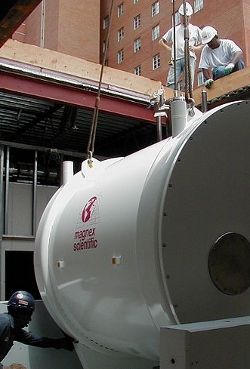
Magnetic resonance imaging (MRI) is a noninvasive, usually painless medical test that helps physicians diagnose and treat medical conditions.
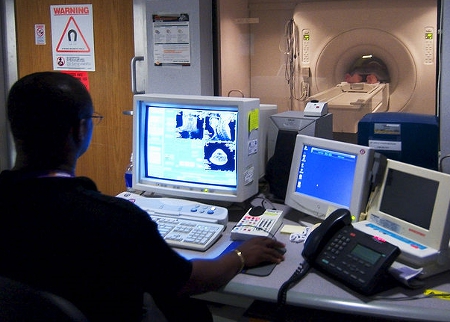
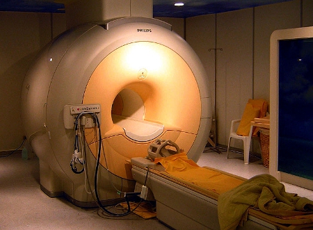
MR imaging uses a powerful magnetic field, radio waves and a computer to produce detailed pictures of organs, soft tissues, bone and virtually all other internal body structures. The images can then be examined on a computer monitor or printed. MRI does not use ionizing radiation (x-rays).
Detailed MR images allow physicians to better evaluate parts of the body and certain diseases that may not be assessed adequately with other imaging methods such as x-ray, ultrasound or computed tomography (also called CT or CAT scanning).
Vocabulary/Definitions
Biomedical Engineering: The application of engineering principles and techniques to the medical field. It combines the design and problem solving skills of engineering with medical and biological sciences to help improve patient health care and the quality of life of individuals.
MRI (Magnetic Resonance Imaging): A noninvasive diagnostic procedure employing an MR scanner to obtain detailed sectional images of the internal structure of the body.
Associated Activities
Visualizing Magnetic Field Lines – Students use a variety of methods to visualize magnetic fields from permanent magnets.
Slinkies and Magnetic Fields – Students use a a metal slinky to mimic the an MRI machine’s magnetic field, running current through the slinky and using computer and calculator software to explore the magnetic field.
PROCEDURE
I. Present the students with the MRI Challenge:
A nearby hospital has just installed a new Magnetic Resonance Imaging facility, which has the capacity to make a three dimensional image of the brain and other parts of the body by putting a patient into a strong magnetic field. The hospital wishes for its entire staff to have a clear knowledge of the risks involved with working near a strong magnetic field, and a basic understanding of why those risks occur. Safety engineers even spend time specifically trying to improve the safety of devices. When an engineer designs a new machine like an MRI machine, he/she must communicate how the design works to the users to ensure they understand how to use it in a safe manner.
Your task over the next few class periods is to educate yourself about the technology of MRI machines, as well as the risks involved, the physics behind those risks, and the safety precautions that should be taken into consideration. At the conclusion of this lesson, you will produce a presentation or pamphlet directed at hospital staff workers that clearly and concisely summarizes MRI technology and safety precautions.
II. Generate ideas: Ask students to work alone to record their personal thoughts and ideas about the problem in their journals. Use one or more of the following journal questions to help students get started:
- What risk factors could a strong field pose to medical personnel?
- How could those risks be reduced or avoided?
- What do you need to know more about?
- What do you already know that is relevant?
Then, after an adequate amount of time, record the students’ thoughts on the board. Try to get ideas from everyone, and write all ideas on the board. If the class is very large, you can have students pair up and share ideas, then get at least one idea from each group.
III. Multiple perspectives: Next, show avideo interview with a medical researcher and the video of removing a chair from an MRI. ( and http://youtube.com/watch?v=FLmKiljrf00) and http://youtube.com/watch?v=4uzJPpC4Wuk.
Allow students to brainstorm additional ideas of needed information. These videos can lead students in new directions and help them understand the uses of MRI.
[youtube]http://youtube.com/watch?v=FLmKiljrf00[/youtube] [youtube]http://youtube.com/watch?v=4uzJPpC4Wuk[/youtube]
Teachers may also assign as homework individual research into recent MRI advances from available news articles and medical or engineering websites, such as this description and video from the West Virginia University on their new “open” MRI.
Finally, break the MRI Challenge down into three knowledge areas that students may want to pursue further in preparing their presentation/phamplet: 1) how magnetic fields affect physical objects, 2) how magnetic fields relate to electricity, and 3) what creates a magnetic field.
IV. Lesson extension — Additional Activity or Writing Exercise
After students are satisfied with their list of needed information and categories, and depending upon the time available, teachers may choose to introduce an associated activity to help students better understand magnetic fields Visualizing Magnetic Field Lines. Otherwise, students should be directed to begin work on their presentations or pamphlets, in-class or as a homework assignment.
V. Lesson assessment
The journal entries students create after being introduced to the MRI Safety Challenge provide opportunities for embedded assessment. The final presentation or pamphlet can also be used to assess their mastery of the material.
Journal Topics:
- What risk factors could a strong field pose to medical personnel?
- How could those risks be reduced or avoided?
- What do you need to know more about?
- What do you already know that is relevant?

Advice for Teachers
This lesson should get students to brainstorm ideas and organize their information. Begin with the students working alone to record their personal thoughts and ideas on the problem. It is helpful to walk around the room and jump-start any students who are drawing a blank. After an adequate amount of time, record the students’ thoughts on the board. Try to solicit ideas from everyone, and write all ideas on the board, overhead projector, or large notepad. If the class is large, you can pair students to share ideas, then get at least one idea from each group.
After the video interview with a real-life researcher and the video of removing a chair from an MRI is viewed, allow students to brainstorm additional ideas of needed information. These videos can lead students in new directions and help them understand the uses of MRI. The goal of this second brainstorming session is to break the MRI Safety Challenge down into three knowledge areas: 1) how magnetic fields affect physical objects, 2) how magnetic fields relate to electricity, and 3) what creates a magnetic field. If students do not raise these topics, you can ask leading questions to help them.
This first section of this lesson allows students to deeply engage with a problem and become motivated to learn information. Section II, Generate Ideas, lets them identify prior knowledge, so they feel that they have a starting place for the problem. Students get to hear everyone’s ideas, so they can learn from one another. This is also a time for teachers to identify and address misconceptions. Section III, Multiple Perspectives, gives students more clues in solving the problem and allows them to see why this information is useful.
References
Dictionary.com. Random House Unabridged Dictionary, Random House. Accessed June 24, 2008. Dictionary.com
Gould, Todd. A. “How MRI Works.” How Stuff Works. How Stuff Works
Hornack, Dr. Joseph P. The Basics of MRI .1996-2007. The Basics of MRI
Radiologyinfo. “MRI of the Body.” Accessed on July 16, 2007. Magnetic Resonance Imaging
Wikipedia.com. Wikimedia Foundation. LastUpdated June 18, 2008. Accessed June 24, 2008. Engineering Design Process
Wikipedia.com. Wikimedia Foundation. LastUpdated June 24, 2008. Accessed June 24, 2008. Magnetic Resonance Imaging
Contributors
Eric Appelt, Primary Author. VU Bioengineering RET Program, School of Engineering, Vanderbilt University
Filed under: Grades 9-12, Lesson Plans
Tags: Biomedical Engineering, Grades 11-12, Safety engineering


